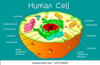
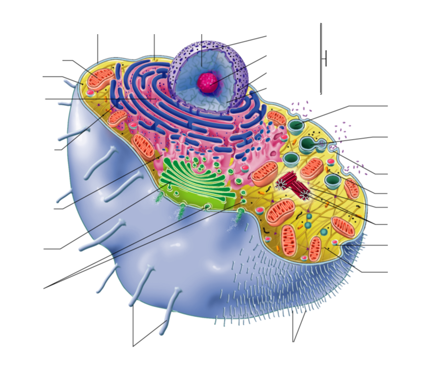
43 diagram of a human cell with labels
PDF Human Cell Diagram, Parts, Pictures, Structure and Functions Human Cell Diagram, Parts, Pictures, Structure and Functions The cell is the basic functional in a human meaning that it is a self-contained and fully operational living entity. Humans are multicellular organisms with various different types of cells that work together to sustain life. Other non-cellular components in the body include water ... Cell: Structure and Functions (With Diagram) - Biology Discussion Cell: Structure and Functions (With Diagram) Let us make an in-depth study of the structure and functions of cell. After reading this article you will learn about: 1. Comparison of Prokaryotic Cells and Eukaryotic Cells and 2. Structure and Components of a Human Cell. Cell is a compartment where all the activities of life takes place.
diagram of a cell labeled Cell cells science inside discover constitution animal structure human anatomy biology body kidsdiscover diagram project peek issues function plant parts Quia - Meiosis Illustration Identification. 18 Pictures about Quia - Meiosis Illustration Identification : A typical cell, labeled diagram. | Alila Medical Images, CELL - Labelled diagram and ...

Diagram of a human cell with labels
Anatomy and Physiology: Parts of a Human Cell - Visible Body The nucleus is a large organelle that contains the cell's genetic information. Most cells have only one nucleus, but some have more than one, and others—like mature red blood cells—don't have one at all. Within the nucleus is a spherical body known as the nucleolus, which contains clusters of protein, DNA, and RNA. cell structure - Pinterest Human Cell coloring page from Anatomy category. Select from 61873 printable crafts of cartoons, nature, animals, Bible and many more. Plant Cell Science Diagram Clipart Set includes: Three diagrams (one labeled, one with blank labels and diagram alone) plus 9 mini-diagrams of different "cell parts." Learn the parts of a cell with diagrams and cell quizzes Two major regions can be found in a cell. The first is the cell nucleus, which houses DNA in the form of chromosomes. The second is the cytoplasm, a thick solution mainly comprised of water, salts, and proteins. The parts of a eukaryotic cell responsible for maintaining cell homeostasis, known as organelles, are located within the cytoplasm.
Diagram of a human cell with labels. The Human Skeleton: All You Need to Know - Bodytomy The human skeletal system functions include providing the body with structure, flexibility and protection. It also helps in the production of red blood cells and white blood cells. It is also a storehouse of minerals and fat tissues. The skeletal system is able to grow very quickly and can adapt to the movement patterns of the body. cell labeled diagram Animal Cells Diagram With Labels Awesome Animal Cell Diagrams Labeled . labeled cells. Spinal Cord Diagram Labeled - Made By Creative Label ... An Annotated Diagram Of A Human Cell . cell human diagram annotated. June 2011 ~ Study Of Biological Science tmfpff.blogspot.com. Animal Cells: Labelled Diagram, Definitions, and Structure - Research Tweet Animal Cells Organelles and Functions. A double layer that supports and protects the cell. Allows materials in and out. The control center of the cell. Nucleus contains majority of cell's the DNA. Popularly known as the "Powerhouse". Breaks down food to produce energy in the form of ATP. File:Diagram human cell nucleus tr.svg - Wikimedia Commons Description. Diagram human cell nucleus tr.svg. en: A diagram of a human cell nucleus, with Turkish labels. Translated version of File:Diagram human cell nucleus.svg, originally created and all rights released by Mariana Ruiz ( User:LadyofHats ). This image is also released to the public domain. az: İnsan hüceyrə nüvəsinin sxematik rəsmi ...
animal bone cell diagram labeled blood microscope under smear peripheral cells labeled histology seen shutterstock illustration lab. Christian Revolution Animal Cell Diagram With Labels And Functions . cell animal diagram labels structure project cells. Connective Tissue - BIOLOGY4ISC biology4isc.weebly.com Circulatory System Labeled Diagram stock illustrations The urinary system. The human urinary system medical illustration with internal organs. Double circulation vector illustration. Labeled educational... Double circulation vector illustration. Labeled educational blood route scheme. Lung capillaries, pulmonary artery, aorta, vein and vena cava diagram. 03 Label the Cell Diagram | Quizlet Start studying 03 Label the Cell. Learn vocabulary, terms, and more with flashcards, games, and other study tools. Label Diagram Human Body Illustrations & Vectors - Dreamstime Download 206 Label Diagram Human Body Stock Illustrations, Vectors & Clipart for FREE or amazingly low rates! New users enjoy 60% OFF. 191,055,749 stock photos online. ... Animal cell structure anatomy infographic diagram. With parts flat vector illustration design for biology science education school book concept microbiology.
Human Cells Printables and Diagrams - The Successful Homeschool Walk your children through the different types of cells found in the human body. These cells include: leukocytes, haematids, thrombocytes, ovum, sperm, sarcomeres, enterocytes, neurons, osteocytes, hepatocytes. They will learn the parts of a cell thanks to a labeled diagram. They will get to see what blood looks like under a microscope without ... Labeled diagram of the human kidney royalty-free images - Shutterstock 189 labeled diagram of the human kidney stock photos, vectors, and illustrations are available royalty-free. See labeled diagram of the human kidney stock video clips. Image type. Structure of Cell: Definition, Types, Diagram, Functions - Embibe What are the five cell structures? Ans: A cell consists of many different structures that have definite shapes, structures, and functions of their own. Some of these structures are (1) Cell Wall (2) Mitochondria (3) Chloroplast (4) Cell Membrane and (5) Nucleus . Q3. What is the structure of a human cell? Cell Organelles- Definition, Structure, Functions, Diagram - Microbe Notes Cell organelles are specialized entities present inside a particular type of cell that performs a specific function. There are various cell organelles, out of which, some are common in most types of cells like cell membranes, nucleus, and cytoplasm. However, some organelles are specific to one particular type of cell-like plastids and cell ...
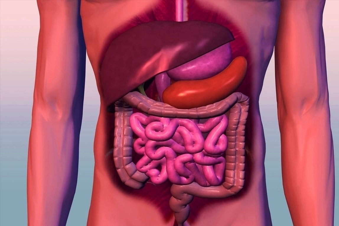
Digestive System | Anatomy System - Human Body Anatomy diagram and chart images
Labelled Diagram Of A Human Cell Bone Cell Labeled Diagram Animal Cell ... Oct 7, 2018 - Labelled Diagram Of A Human Cell Bone Cell Labeled Diagram Animal Cell Free Printable To Label. Oct 7, 2018 - Labelled Diagram Of A Human Cell Bone Cell Labeled Diagram Animal Cell Free Printable To Label. Pinterest. Today. Explore. When autocomplete results are available use up and down arrows to review and enter to select. Touch ...

JQ Nursing Review: A&P Lecture 1: The Cell
Human Heart Diagram Labeled | Science Trends The human heart is an organ responsible for pumping blood through the body, moving the blood (which carries valuable oxygen) to all the tissues in the body. Without the heart, the tissues couldn't get the oxygen they need and would die. Along with lymphatic vessels, the blood, blood vessels, and lymph, the heart composes the circulatory ...

Cell Biology – Cells, Tissues, Organs & Systems | Human cell structure, Cell diagram, Cell structure
Cells Diagram | Science Illustration Solutions - Edrawsoft Cells Diagram. Cells are the basic building blocks of all living things. The human body is composed of trillions of cells. Cells have many parts, each with a different function. Some of these parts, called organelles, are specialized structures that perform certain tasks within the cell. Drawing cells diagram helps you better understand your ...
Animal Cell Images, Stock Photos & Vectors | Shutterstock
Labeled Plant Cell With Diagrams | Science Trends Plant cells contain many organelles such as ribosomes, the nucleus, the plasma membrane, the cell wall, mitochondria, and chloroplasts. In addition, plant cells differ from animal cells in a number of key ways. Examining a diagram of the plant cell will help make the differences clearer. Let's go over the individual components of plant cells ...
7 Best Images of Neuron Label Worksheet - Blank Neuron Cell Diagram, Synapse Neuron Worksheet ...
A Labeled Diagram of the Animal Cell and its Organelles As observed in the labeled animal cell diagram, the cell membrane forms the confining factor of the cell, that is it envelopes the cell constituents together and gives the cell its shape, form, and existence. ... it is essential that the DNA remains intact and gets evenly distributed among the cells. Every human body cell contains 46 ...

An Annotated Diagram Of A Human Cell
Blood Cell Diagram Pictures, Images and Stock Photos Blood stem cell is an immature cell that can develop into all types of blood cells, including white blood cells, red blood cells, and platelets. Blood stem cells are found in the peripheral blood and the bone marrow. Also called hematopoietic stem cell. 3d render. Multiple myeloma. plasma cell myeloma.

Cell Review Guide Answers in 2020 | Human cell structure, Human cell diagram, Animal cell structure
Human Cell Organelles Labeling Diagram | Quizlet Start studying Human Cell Organelles Labeling. Learn vocabulary, terms, and more with flashcards, games, and other study tools.

Biology for Kids: Cells, Organisms, and the Diversity of Life | Human cell diagram, Human cell ...
Skeletal System - Labeled Diagrams of the Human Skeleton - Innerbody The skeletal system includes all of the bones and joints in the body. Each bone is a complex living organ that is made up of many cells, protein fibers, and minerals. The skeleton acts as a scaffold by providing support and protection for the soft tissues that make up the rest of the body. The skeletal system also provides attachment points for ...

Animal Cell Model: What To Use - YouTube
Labeled Diagram of the Human Kidney - Bodytomy Size of an adult kidney: Length: 11-12 cm. Width: 5.0-7.5 cm. Weight of an adult kidney: Males: 125-170 g. Females: 115-155 g. Located in the abdominal cavity, kidneys are the most efficient filters. They are an important component of the human excretory system, and help the body retain essential molecules and get rid of the unwanted ones.
Human Cell Diagram Labeled - Diagram Media
Cell Diagram | Free Cell Diagram Templates - Edrawsoft A free customizable cells diagram template is provided to download and print. Quickly get a head-start when creating your own cell diagram. Here is a simple cell diagram example created by Science Diagram Maker.
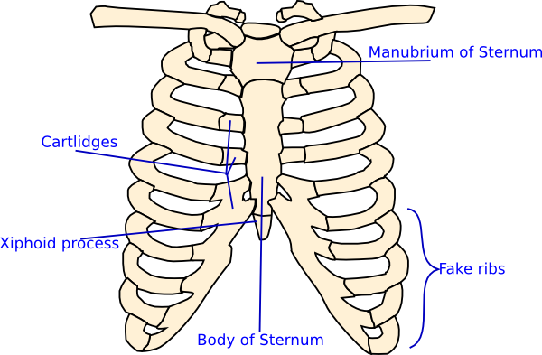
Labelled Rib Cage Clip Art at Clker.com - vector clip art online, royalty free & public domain
Liver Diagram with Detailed Illustrations and Clear Labels - BYJUS Liver Diagram. The liver is one of the most important organs in the human body. Anatomically, the liver is a meaty organ that consists of two large sections called the right and the left lobe. The rib cage partly protects the liver and cannot be felt if you were to touch it. However, it can be felt ascending and descending if you were to take a ...

Biology Unit: Cells, Organs, Systems - Ms. Corner Gardiner's Classes
Learn the parts of a cell with diagrams and cell quizzes Two major regions can be found in a cell. The first is the cell nucleus, which houses DNA in the form of chromosomes. The second is the cytoplasm, a thick solution mainly comprised of water, salts, and proteins. The parts of a eukaryotic cell responsible for maintaining cell homeostasis, known as organelles, are located within the cytoplasm.

Questions And Answers On Labeled/Unlebled Diagrams Of A Human Cell / Question 14: Draw a labeled ...
cell structure - Pinterest Human Cell coloring page from Anatomy category. Select from 61873 printable crafts of cartoons, nature, animals, Bible and many more. Plant Cell Science Diagram Clipart Set includes: Three diagrams (one labeled, one with blank labels and diagram alone) plus 9 mini-diagrams of different "cell parts."

The Human Egg Cell Explained For Egg Donors - Altrui Egg Donation Agency
Anatomy and Physiology: Parts of a Human Cell - Visible Body The nucleus is a large organelle that contains the cell's genetic information. Most cells have only one nucleus, but some have more than one, and others—like mature red blood cells—don't have one at all. Within the nucleus is a spherical body known as the nucleolus, which contains clusters of protein, DNA, and RNA.
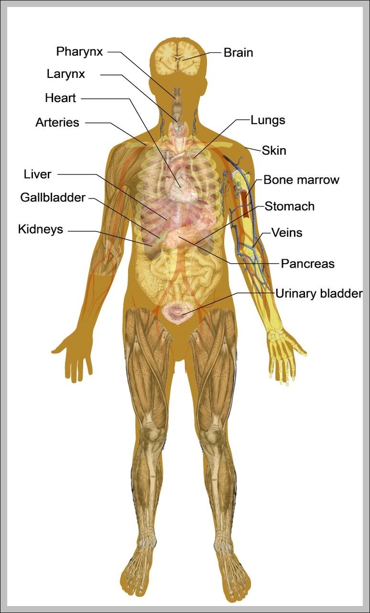
anatomy of the male body 744×1293 | Anatomy System - Human Body Anatomy diagram and chart images
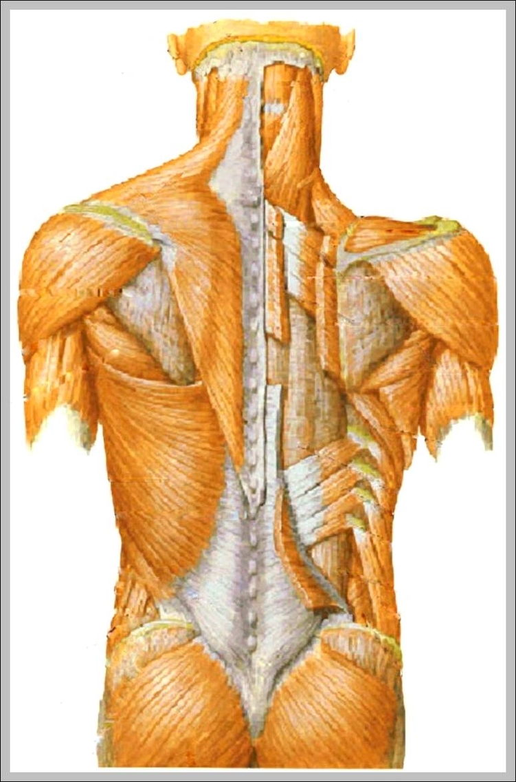
muscular pictures 744×1179 | Anatomy System - Human Body Anatomy diagram and chart images
Post a Comment for "43 diagram of a human cell with labels"